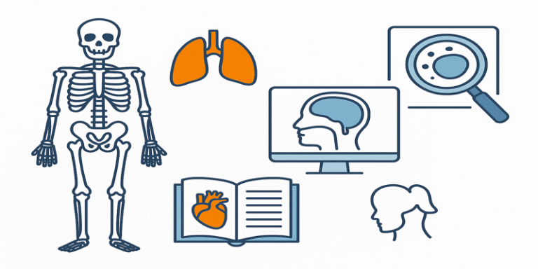
Anatomy is one of the oldest and most fundamental branches of the medical sciences, devoted to the study of the structure of the human body. It encompasses everything from the macroscopic organisation of organs and body systems to the microscopic architecture of cells and tissues. Anatomical knowledge has been the foundation upon which modern medicine has been built, allowing practitioners to identify diseases, perform surgeries, and interpret diagnostic images with precision.
As Standring (2020) notes, accurate anatomical understanding is critical for healthcare professionals, whether they are diagnosing a condition, navigating a complex surgical procedure, or developing new medical technologies.
1.0 Divisions of Anatomy
Anatomy is typically divided into several major branches, each focusing on different levels of structural organisation.
1.1 Gross Anatomy
Gross anatomy (or macroscopic anatomy) deals with structures that can be seen with the naked eye. It is traditionally studied through dissection of cadavers, allowing students and medical practitioners to gain a three-dimensional understanding of the body’s organisation. Gross anatomy can be approached in two main ways:
- Regional anatomy: Studying all structures within a specific body region, such as the head and neck or the thorax.
- Systemic anatomy: Studying body systems individually, such as the skeletal system, muscular system, or nervous system.
Gross anatomy provides the foundation for surgical practice, as surgeons must visualise the spatial relationships between structures to operate safely.
1.2 Microscopic Anatomy (Histology)
Microscopic anatomy, also called histology, focuses on cells and tissues that require magnification for observation. Using light microscopes and electron microscopes, histologists examine the detailed structures of tissues, identifying their organisation and specialisation.
Histology is essential for diagnosing many diseases. For example, a biopsy taken from a suspicious lump can be examined histologically to confirm whether it is benign or malignant (Junquiera & Carneiro, 2015).
1.3 Developmental Anatomy (Embryology)
Developmental anatomy studies how structures form and change from conception to adulthood. This includes:
- Embryology: The study of the first eight weeks of development.
- Foetal development: Growth and maturation until birth.
- Postnatal changes: Structural adaptations throughout life.
Embryology is particularly important for understanding congenital disorders and structural malformations.
1.4 Comparative Anatomy
Comparative anatomy examines structural similarities and differences between human anatomy and that of other species. This field helps in understanding evolutionary relationships and in using animal models for medical research.
2.0 Importance of Anatomy in Medicine
2.1 Surgery
For surgeons, precise anatomical knowledge is not optional—it is life-saving. During operations, they must navigate blood vessels, nerves, and organs without causing unnecessary damage. For example, knowledge of the Circle of Willis in the brain is crucial in neurosurgery to prevent life-threatening complications.
2.2 Diagnostic Imaging
Anatomy forms the basis for interpreting X-rays, MRI scans, CT scans, and ultrasound images. Radiologists use their knowledge of normal anatomical structures to identify abnormalities, such as tumours, fractures, or organ enlargement.
2.3 Physical Examination
Doctors performing a physical exam rely on anatomical landmarks to guide their assessment. Palpating the abdomen or checking the pulse in the radial artery both require knowledge of surface anatomy.
3.0 Anatomy and Education
Anatomy is a core component of medical, nursing, physiotherapy, and sports science curricula. The teaching of anatomy often combines:
- Cadaver dissection for hands-on experience.
- Prosection (studying pre-dissected specimens) for focused learning.
- Medical imaging to visualise anatomy in living patients.
- 3D computer models and virtual reality simulations for interactive learning.
Recent research (Estai & Bunt, 2016) has highlighted that combining traditional dissection with digital tools enhances student understanding and retention.
4.0 Modern Advances in Anatomical Science
4.1 Imaging Technologies
Advances in MRI and CT scanning have revolutionised anatomy by allowing detailed visualisation of structures in living patients. Functional MRI (fMRI) goes further, showing which areas of the brain are active during specific tasks.
4.2 Virtual and Augmented Reality
Medical schools now use virtual reality (VR) and augmented reality (AR) to teach anatomy. These tools allow students to interact with life-sized 3D models, providing an immersive learning experience.
4.3 Plastination
Plastination, developed by Gunther von Hagens, preserves body tissues using polymers. This technique produces durable anatomical specimens for study without the health risks associated with formaldehyde.
5.0 Challenges in Learning Anatomy
Despite its importance, students often find anatomy challenging due to:
- The volume of information to memorise.
- The need to understand three-dimensional relationships.
- Variations in anatomy between individuals.
Effective learning requires a combination of visual, tactile, and applied methods (Sugand et al., 2010).
6.0 Anatomy Beyond Medicine
While anatomy is central to healthcare, it also has applications in other fields:
- Forensic science: Determining cause of death or identifying remains.
- Sports science: Improving athletic performance by understanding muscle and joint mechanics.
- Anthropology: Studying human evolution and variation.
7.0 Ethical Considerations
Anatomy has a complex ethical history, particularly regarding the use of human bodies for dissection. Today, medical institutions operate under strict ethical guidelines, with donated bodies treated with dignity and respect (Jones, 2014).
8.0 The Future of Anatomy
Emerging trends point to even greater integration of digital technology and artificial intelligence in anatomical studies. AI can assist in identifying structures in medical imaging, while 3D bioprinting may allow for the creation of lifelike anatomical models for surgical training.
Anatomy remains the bedrock of medical science, linking the study of structure to the practice of healing. From the cadaver lab to the operating theatre, and from the lecture hall to the research lab, anatomy informs every aspect of healthcare. As technology continues to advance, the way we study and apply anatomical knowledge will evolve, but its fundamental importance will remain unchanged.
Whether guiding a surgeon’s scalpel, helping a physiotherapist design a rehabilitation plan, or enabling a forensic scientist to solve a mystery, anatomy is the silent framework supporting human health and understanding.
References
Estai, M. & Bunt, S. (2016). Best teaching practices in anatomy education: A critical review. Annals of Anatomy, 208, 151–157.
Jones, D. G. (2014). Ethical issues in anatomy: The impact of recent history on modern practice. Clinical Anatomy, 27(3), 304–313.
Junquiera, L. C. & Carneiro, J. (2015). Basic Histology: Text and Atlas (13th ed.). McGraw-Hill Education.
Standring, S. (2020). Gray’s Anatomy: The Anatomical Basis of Clinical Practice (42nd ed.). Elsevier.
Sugand, K., Abrahams, P. & Khurana, A. (2010). The anatomy of anatomy: A review for its modernization. Anatomical Sciences Education, 3(2), 83–93.